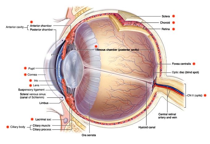
- Anterior Cavity
- Anterior Chamber: Fluid filled space between the iris and the innter layer of the cornea
- Posterior Chamber: Small space between the irin and the lens
- Pupil: The black part of the eye; the pupil is an opening in the iris that allows light into the interior of the eye and the retina. The size of the pupil dilates or contracts in response to the amount of light. In humans, pupils are round, but pupils in other animals vary in shape.
- Cornea: The cornea consists of six layers, and covers the iris, pupil, and anterior chamber of the eye. It is responsible for the majority of the eye’s ability to refract light, although it is not able to adjust focus. Contact lenses are designed to sit on top of the cornea, and proper fitting of contact lenses requires measuring the exact size and curvature of your cornea.
- Iris: The iris surrounds the pupil and regulates its size in response to light. The iris is the colored part of the eye, which can vary based on genetics.
- Lens: The lens, along with the cornea, is responsible for refracting light entering the eye. The lens changes shape to adjust the focal distance, allowing us to view images at various distances. This ability is known as accomodation. Over time, the lens loses its ability to focus on close-up objects (a condition known as presbyopia). This usually happens around age 40-45, after which most people begin to require reading glasses or multifocal lenses.
- Suspensory Ligament: Suspends the lens in palce
- Scleral Venous Sinus (Canal of Schlemm): Drains aqueous humor from the interior of the eye. If blocked, intra-ocular pressure can increase, leading to glaucoma.
- Limbus: Connects the cornea to the sclera.
- Lacrimal Sac: Collects tears, bacteria, and debris.
- Ciliary Body
- Ciliary Muscle: Ring of muscle that controls accommodation by chanign the shape of the lens.
- Ciliary Process: Ciliary processes are responsible for the production of aqueous humor.
- Ora Serrata: Junction between the retina and ciliary body.
- Sclera: aka the “white” of the eye. The sclera helps maintain the shape of the eye, and provides protection from internal and external pressure.
- Choroid: The choroid is a layer of connective tissure between the retina and sclera that provides nutrients to the retina.
- Retina: Consists of layers of neurons and photoreceptor cells responsible for visual perception. The retina is connected to the brain via the optic nerve. The cornea and lens are responsible for focusing light on the retina. Myopia (nearsightedness) is a condition where light focuses in front of the retina rather than directly on it. Hyperopia (farsightedness) is a condition where light focuses behind the retina. Presbyopia is when the lens cannot accommodate to focus close-up objects (such as when reading). All of these conditions and more can be corrected to some degree with eyeglasses and/or contact lenses, which change the focal point of the light entering the eye. Other treatment options to correct refractive conditions can include refractive surgery and orthokeratology (the use of a rigid contact lens designed to reshape the cornea).
- Vitreous Chamber: Contains vitrous humor, a transparent gel that fills the interior of the eye. The vitreous is connected to the Ora serrata.
- Fovea Centralis: The fovea is located in the center of the macula, and is responsible for central vision
- Optic Disc (Blind Spot): The optic disc is the region of the eye where the optic nerve forms and connects to the brain. It is known as the “blind spot” due to the lack of photoreceptor cells.
- CN II (Optic): The optic nerve, also known as crianial nerve 2, transmits information from the retina to the brain. Glaucoma is characterized by optic nerve damage resulting in visual field loss.
- Central Retinal Artery/Vein: Provides blood flow to and from the ocular region.
- Hyaloid Canal: Transparent canal running through the vitreous from the optic nerve to the lens.